Bone Cross Section Microscope - Cross Section Of Spinal Cord Under The Microscope View ... - Cross section performed on focused electon beam (fib) microscope at the university of kentucky's electron microscopy center.
Bone Cross Section Microscope - Cross Section Of Spinal Cord Under The Microscope View ... - Cross section performed on focused electon beam (fib) microscope at the university of kentucky's electron microscopy center.. The finished bone section will be bonded to a microscope slide and so the first step is to grind flat and polish the part of the bone that will be glued to the slide. Thus as usual microscopic cross sections are experimentally measured. Accuracy of the tested digitization method was expressed by. Jump to navigation jump to search. Structural parts of a microscope and their functions.
From wikimedia commons, the free media repository. Use electromagnets to focus electrons resulting in significantly greater magnifications and resolutions. Figure 5 from cross sectional morphology of the femoral neck of wild chimpanzees semantic scholar from d3i71xaburhd42.cloudfront.net. 1, cmp consists of both crystalline and glass phases fig. Sometimes referred to as 'spongy bone' or 'trabecular bone', cancellous bone is found within the middle of large bones.
Pin by Danielle Papas on Nursing Anatomy & Physiology ... from i.pinimg.com Bone marrow aspiration uses a hollow needle to remove a small sample (about 1 ml) of bone marrow for examination under a microscope. Sometimes referred to as 'spongy bone' or 'trabecular bone', cancellous bone is found within the middle of large bones. Cross section performed on focused electon beam (fib) microscope at the university of kentucky's electron microscopy center. See more ideas about microscopic, plant cell, microscopic. They have stage clips hold the specimen slides in place. A cross section of a human long bone. A cross section of a compact bone shows concentric circles called lamellae. Structural parts of a microscope and their functions.
Important features in the bone cross section such as harvesian canals, osteons, osteon fragments, lamellar bone, bony trabeculae, myxoid matrix and artifact for.
Bone marrow aspiration uses a hollow needle to remove a small sample (about 1 ml) of bone marrow for examination under a microscope. These bone cells have long branching arms (d) which lets them communicate with. This simply involves placing a section of the bone on the microscope stage and viewing. A cross section of a compact bone shows concentric circles called lamellae. From wikimedia commons, the free media repository. Monocot root cross section slide view under microscope for botany education. Thin section of dinosaur bone. Overview of microscope and diagram. In the last decade, considerable technological improvements have been made to repair damaged bones and tissue, such as bone cross sections with implants for microscopic examinations. Jump to navigation jump to search. They build the entire picture, improve your understanding, consolidate the information and facilitate recall. When the light that enters the condenser is polarized by placing a polarizer in the filter holder and a second, crossed polarizer at the image plane. Thus as usual microscopic cross sections are experimentally measured.
They build the entire picture, improve your understanding, consolidate the information and facilitate recall. Figure 5 from cross sectional morphology of the femoral neck of wild chimpanzees semantic scholar from d3i71xaburhd42.cloudfront.net. Jump to navigation jump to search. Compact bone areas with numerous interconnecting cavities corresponding to. Both types of bone marrow are enriched with blood vessels and capillaries.2.
A New Probe for CSI? | ArcNews from www.esri.com The finished bone section will be bonded to a microscope slide and so the first step is to grind flat and polish the part of the bone that will be glued to the slide. Jump to navigation jump to search. The nuclear cross section of a nucleus is used to describe the probability that a nuclear reaction will occur. Accuracy of the tested digitization method was expressed by. The large dark spots are passages for blood vessels and nerves. Hope you enjoy and please. See more ideas about microscopic, plant cell, microscopic. Both types of bone marrow are enriched with blood vessels and capillaries.2.
This simply involves placing a section of the bone on the microscope stage and viewing.
Important features in the bone cross section such as harvesian canals, osteons, osteon fragments, lamellar bone, bony trabeculae, myxoid matrix and artifact for. Hope you enjoy and please. In this short video i use blender 2.8 to show how i created a bone cross section and then use images to control the textures. A cross section of a compact bone shows concentric circles called lamellae. New vs old newly designed microscope slide for cutting and viewing a quick cross section of textile fibers and small soft specimens of many types. Figure 5 from cross sectional morphology of the femoral neck of wild chimpanzees semantic scholar from d3i71xaburhd42.cloudfront.net. Overview of microscope and diagram. 1, cmp consists of both crystalline and glass phases fig. File:earthworm crosssection stained microscope slide labeled.jpg. The microscopic cross section measures the probability of occurrence of a particular nuclear reaction. Cross section performed on focused electon beam (fib) microscope at the university of kentucky's electron microscopy center. A cross section of a human long bone. These bone cells have long branching arms (d) which lets them communicate with.
Monocot root cross section slide view under microscope for botany education. Thus as usual microscopic cross sections are experimentally measured. Cross section performed on focused electon beam (fib) microscope at the university of kentucky's electron microscopy center. The microscopic cross section measures the probability of occurrence of a particular nuclear reaction. Monocots and dicots have distinct vascular bundles organization.
Molecular Expressions Microscopy Primer: Specialized ... from micro.magnet.fsu.edu Use electromagnets to focus electrons resulting in significantly greater magnifications and resolutions. Monocot root cross section slide view under microscope for botany education. From wikimedia commons, the free media repository. The nuclear cross section of a nucleus is used to describe the probability that a nuclear reaction will occur. The microscopic cross section measures the probability of occurrence of a particular nuclear reaction. Microscope cross section (page 1). A cross section of a compact bone shows concentric circles called lamellae. Bone marrow aspiration uses a hollow needle to remove a small sample (about 1 ml) of bone marrow for examination under a microscope.
Bone marrow aspiration uses a hollow needle to remove a small sample (about 1 ml) of bone marrow for examination under a microscope.
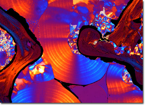
From wikimedia commons, the free media repository. Important features in the bone cross section such as harvesian canals, osteons, osteon fragments, lamellar bone, bony trabeculae, myxoid matrix and artifact for. In the last decade, considerable technological improvements have been made to repair damaged bones and tissue, such as bone cross sections with implants for microscopic examinations. Hope you enjoy and please. In this short video i use blender 2.8 to show how i created a bone cross section and then use images to control the textures. Sometimes referred to as 'spongy bone' or 'trabecular bone', cancellous bone is found within the middle of large bones. Cross section of ground compact bone. Use electromagnets to focus electrons resulting in significantly greater magnifications and resolutions. This simply involves placing a section of the bone on the microscope stage and viewing. Thin section of dinosaur bone. The microscopic bone cross section image acquired by using electronic microscope and is shown in fig.2. Bone marrow aspiration uses a hollow needle to remove a small sample (about 1 ml) of bone marrow for examination under a microscope. See more ideas about microscopic, plant cell, microscopic.
Hope you enjoy and please bone cross section. Accuracy of the tested digitization method was expressed by.


0 Komentar